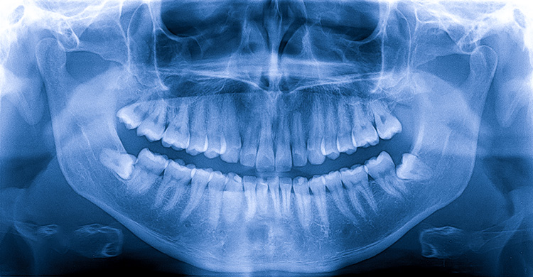
Dental X-Ray
X-rays, also known as radiographs, are an essential part of any dental care treatment plan. They are diagnostic, but they can also be preventative, by helping a dentist diagnose potential oral care issues in a patient’s mouth before they become a major problem. An x-ray is a type of energy that passes through soft tissues and is absorbed by dense tissue. Teeth and bone are very dense, so they absorb X-rays, while X-rays pass more easily through gums and cheeks.
X-rays are divided into two main categories, intraoral and extraoral. Intraoral is an X-ray that is taken inside the mouth. An extraoral X-ray is taken outside of the mouth.
Intraoral X-rays are the most common type of radiograph taken in dentistry. They give a high level of detail of the tooth, bone and supporting tissues of the mouth. These X-rays allow dentists to :
Services Provided :
Oral Medicine & Radiologists :


